
| name | Amanita pachyvolvata |
| name status | nomen acceptum |
| author | (Bon) Krieglst. |
| english name | "Bon's Great Ringless Amanita" |
| cap | The cap of Amanita pachyvolvata is 50 - 80 (-110) mm wide, hemispheric or ovate, then plano-convex, shiny or subviscid, rather pruinose toward the margin, more mat toward the center, with a striate-sulcate margin (35±% of the radius), becoming rimose. The cap is sordid ochraceous or fauve-gray to bister over disc and is a paler brown-yellow toward margin. No remains of the volva are present on the cap. |
| gills | The gills are creamy white or white or yellowish white, rather broad, with a fimbriate edge or slightly fimbriate and serrate edge. No information is available to the authors regarding short gills in this species. |
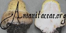
| stem | The stem is (120-) 150 - 200 × 20 - 30 mm or (120-) 150 - 180 (-200) × (10-) 15 - 30 mm, white, then more or less grayish or yellowish gray, variegated, narrowing upward, and without a ring. The flesh is stuffed at first, then quickly hollow. The very large volval sac is white, membranous, and persistent. |
| spores | The spores measure (10.2-) 11.0 - 14.2 (-15.0) × (9.5-) 10.8 - 13.2 (-15.0) µm and are globose to subglobose to broadly ellipsoid and inamyloid. Clamps are infrequent at bases of basidia. |
| discussion |
This species was originally described from France (Dép. Haute-Loire) under Fir and Spruce. For pages concerning similar species, see A. pachycolea D. E. Stuntz in Thiers & Ammirati, A. magnivolvata Aalto, A. praelongipes Kärcher & Contu, and Amanita violettae Tulloss.—R. E. Tulloss |
| brief editors | RET |
| name | Amanita pachyvolvata | ||||||||
| author | (Bon) Krieglst. 1984. Beih. Z. Mykol. 5: 191, fig. opposite p. 190. | ||||||||
| name status | nomen acceptum | ||||||||
| english name | "Bon's Great Ringless Amanita" | ||||||||
| synonyms |
≡Amanitopsis pachyvolvata Bon. 1978. Doc. Mycol. 8(29): 36.
≡Amanita pachyvolvata (Bon) Garcin nom. inval. 1984. Amanites Européennes: 165. [Autographic publication, etc. ICBN §29.3, §29.4, §32.1a.]
≡Amanita pachyvolvata (Bon) Contu. 1985c. Funghi Ambiente 3(=no. 40): 24. [Superfluous combination.]
≡Amanita pachyvolvata (Bon) D. A. Reid. 1987. Notes Roy. Bot. Gard. Edinburgh 44: 511. [Superfluous combination.]
≡Amanita pachyvolvata (Bon) Romagn. nom. inval. 1992. Bull. Trimestriel Soc. Mycol. France 108: 73. [Lacking full and direct reference to basionym. Superfluous combination.] The editors of this site owe a great debt to Dr. Cornelis Bas whose famous cigar box files of Amanita nomenclatural information gathered over three or more decades were made available to RET for computerization and make up the lion's share of the nomenclatural information presented on this site. | ||||||||
| etymology | παχυς, thick + volvatus, having a volva | ||||||||
| MycoBank nos. | 105913, 308605 | ||||||||
| GenBank nos. |
Due to delays in data processing at GenBank, some accession numbers may lead to unreleased (pending) pages.
These pages will eventually be made live, so try again later.
| ||||||||
| holotypes | in herb. M. Bon => LIP | ||||||||
| type studies | Tulloss. 1994. Mycotaxon 52: 366, figs. 37-38. | ||||||||
| selected illustrations | Bon. 1979. Fungorum Rar. Icon. Color. 11: 37, fig. 6g-i [microscopy] and pl. 88. | ||||||||
| intro |
The following text may make multiple use of each data field. The field may contain magenta text representing a type study or a study of original material by Tulloss. The same field may also contain black text, which will represent a revision of the species by Tulloss. Paragraphs of black text will be labeled if further subdivision of this text is appropriate. Olive text indicates a specimen that has not been thoroughly examined (for example, for microscopic details) and marks other places in the text where data is missing or uncertain. The macroscopic data below is based on the protolog and Bon's illustrated revision (1979). The microscopic data of the following is based upon original research by R. E. Tulloss (1994). | ||||||||
| pileus | Tulloss (1994): 50 - 80 (-110) mm wide, sordid ochraceous or fauve-gray to bister over disc and a paler brown-yellow (with olive tints in early stages of expansion) toward margin, hemispheric or ovate, then plano-convex, shiny or subviscid, rather pruinose toward margin, more mat toward disc, with disc flecked or rivulose due to concentrically arranged, beige or somewhat brownish, fine squamules; context white; margin striate-sulcate (0.35±R), straight, becoming rimose; universal veil absent. | ||||||||
| lamellae | Tulloss (1994): creamy white or white, rather broad, "segmentiform" (Bon, 1979), with edge fimbriate or slightly fimbriate and serrate (Bon, 1979) and yellowish white; lamellulae not described. | ||||||||
| stipe | Tulloss (1994): (120-) 150 - 200 × 20 - 30 mm (Bon, 1978) or (120-) 150 - 180 (-200) × (10-) 15 - 30 mm (Bon, 1979), white, then more or less grayish (Bon, 1978) or yellowish gray (Bon, 1979), variegated, narrowing upward; context stuffed at first, then quickly hollow, with central cylinder often having diameter greater than half of that of stipe; exannulate; universal veil as a very large volval sac, white, membranous, persistent, 4 - 7 mm (Bon, 1978) or 4 - 6 mm (Bon, 1979) thick, ovoid at first and retaining a subcylindric to elongately ellipsoid form for some time, with overall dimensions 60 - 80 (-100) × 30 - 50 (-60) mm, obtuse below, smooth to slightly furfuraceous on exterior, not bruising or staining except sometimes more or less ochraceous toward base in older material, with low, rounded limbus internus at point of juncture between stipe and volval limb. | ||||||||
| odor/taste | Tulloss (1994): neither recorded. | ||||||||
| macrochemical tests |
Tulloss (1994): none recorded. | ||||||||
| pileipellis | Tulloss (1994): 100 - 140 µm thick, yellow-brown to orange-brown intracellular pigment mostly concentrated in hyphae of lower half; filamentous, undifferentiated hyphae 1.0 - 6.7 µm wide, subradially arranged, partially to nearly completely gelatinizing at surface, densely packed vertically; refractive hyphae 2.8 - 6.0 µm wide, serpentine. | ||||||||
| pileus context | Tulloss (1994): filamentous, undifferentiated hyphae 1.8 - 9.5 µm wide, frequently branching, in fascicles that are loosely interwoven; acrophysalides terminal, singly or in short chain, thin-walled, subcylindric to clavate to ovoid to subpyriform, to 70 × 42 µm; refractive hyphae 2.8 - 11.6 µm wide, branching, common, locally loosely knotted, at times penetrating pileipellis. | ||||||||
| lamella trama | Tulloss (1994): obscurely bilateral (divergent structures close to base of basidia) [probably poorly rehydrate tissues, needs revision in better material]; wcs = 85 - 110± µm in immature specimen; filamentous, undifferentiated hyphae 1.8 - 11.2 µm wide, dominating and obscuring other structures, branching, with those of higher diameter having constrictions at septa and intergrading with chains of subventricose to ellipsoid(?) segments (up to 61 × 33 µm); terminal, inflated cells not observed; refractive hyphae 3.2 - 5.8 µm wide. | ||||||||
| subhymenium |
Tulloss (1994): wst-near = (0-) 5± µm and wst-far = 25± (-40) µm in immature specimen; basidia arising from thin layer (1 - 3 cells thick) of small cells (subglobose to ovoid to clavate) and from partially inflated and uninflated hyphal segments (latter often perpendicular to hymenial surface, but arising from hyphae running parallel to that surface with only one or two intervening, short, intercalary segments; filamentous, undifferentiated hyphae running parallel to hymenial surface commonly seen in subhymenial region. In immature material, inflated cells somewhat fewer, but otherwise above description holding. In mature material, subhymenium and basidia largely destroyed due to infection by hyphomycete with thick-walled, conidia-bearing hyphae. [Note: The above is an inadequate description due to the quality of the collection when it was revised.—ed.] | ||||||||
| basidia |
Tulloss (1994): 55 - 66 × 14.5 - 21 µm, 4- and, occasionally, 1-sterigmate, thin-walled; clamps present [less frequent(?) in more mature material]. [Note: The range of lengths of basidia is suspiciously limited. Revision of well-dried, mature material is needed.—ed.] | ||||||||
| universal veil | Tulloss (1994): On pileus: absent. On stipe base, exterior surface: outermost layer (only 2 - 3 hyphal diameters thick) consists of very loosely interwoven fascicles of slightly gelatinizing to gelatinizing filamentous, undifferentiated hyphae, with majority having very narrow diameter, with hyphae below this layer forming tighter weave; filamentous, undifferentiated hyphae 1.8 - 7.4 (-15.4) µm wide, branching, tip cells occasionally with small particles on very tip; refractive hyphae 1.8 - 7.0 µm wide, branching, locally common. On stipe base, interior: filamentous, undifferentiated hyphae 3.5 - 14.4 µm wide, loosely interwoven, loosely coiling, branching, dominant; inflated cells terminal, thin-walled, plentiful, subcylindric to narrowly clavate to clavate to ellipsoid to subpyriform, up to 105 × 70 µm; refractive hyphae 3.5 - 6.7 µm wide. On stipe base inner surface: like interior, but slightly gelatinized. | ||||||||
| stipe context | Tulloss (1994): longitudinally acrophysalidic; filamentous, undifferentiated hyphae 1.8 - 6.0 µm wide; acrophysalides thin-walled, plentiful, up to 219 × 40 µm, most smaller than 155 µm long; refractive hyphae 4.5 - 5.2 µm wide, branching. | ||||||||
| partial veil | Tulloss (1994): absent. | ||||||||
| lamella edge tissue |
Tulloss (1994): not described. sterile. | ||||||||
| basidiospores | Tulloss (1994): [40/1/1] (10.2-) 11.0 - 14.2 (-15.0) × (9.5-) 10.8 - 13.2 (-15.0) µm, (L = 12.4 µm; W = 11.5 µm; Q = (1.0-) 1.02 - 1.14 (-1.20); Q = 1.08), inamyloid, smooth, thin-walled, hyaline, colorless, globose to subglobose to (infrequently) broadly ellipsoid, often adaxially flattened; apiculus sublateral, prominent, cylindric; contents mono- or biguttulate; color in deposit not recorded. | ||||||||
| ecology | Tulloss (1994): Under Picea and Abies, by edge of road. | ||||||||
| material examined | Tulloss (1994): FRANCE: HAUTE-LOIRE—Le Puy, Lac du Bouchet, 6.ix.1977 M. Bon 770906 (holotype, in herb. M. Bon => LIP). | ||||||||
| discussion |
The packet of holotype material loaned to me contained an immature basidiome in which the pileus was just free of the tubular, saccate volva. Sporulation seems not to have begun in this specimen, which unfortunately makes up most of the material I have reviewed. The packet contained a few narrow, radial slices of pileus with mature lamellae attached; plentiful spores were found on these fragments; however, the mature structure of the lamellae was not clearly discernible due to destruction caused by a mold. The dimensions given above for the lamella trama and subhymenium may be less than would be found in a mature, well-preserved specimen. Acrophysalides of the stipe context may not be fully inflated in the immature material examined. When I borrowed this collection a second time, it arrived badly broken in the mails. Fortunately, I had drawn the stipe base of the immature specimen including the surrounding volval sac when I first borrowed the material. I have deposited that drawing with the collection. For comparison with A. magnivolata, A. pachycolea, and A. violettae, see the discussions following the descriptions of those species. | ||||||||
| citations | —R. E. Tulloss | ||||||||
| editors | RET | ||||||||
Information to support the viewer in reading the content of "technical" tabs can be found here.
Each spore data set is intended to comprise a set of measurements from a single specimen made by a single observer; and explanations prepared for this site talk about specimen-observer pairs associated with each data set. Combining more data into a single data set is non-optimal because it obscures observer differences (which may be valuable for instructional purposes, for example) and may obscure instances in which a single collection inadvertently contains a mixture of taxa.


Text and User-Generated Sporographs are published under the Creative Commons License.

In the case of a taxon page, image credits are on the 'image' tab.